X-Ray Phase-Contrast Radiography of Cavitation Bubbles
Laser-induced cavitation bubbles collapsing near a solid boundary are investigated using the time-resolved phase-contrast capabilities offered by the powerful ESRF synchrotron x-ray source to capture detailed hidden features of the bubble interface.
-
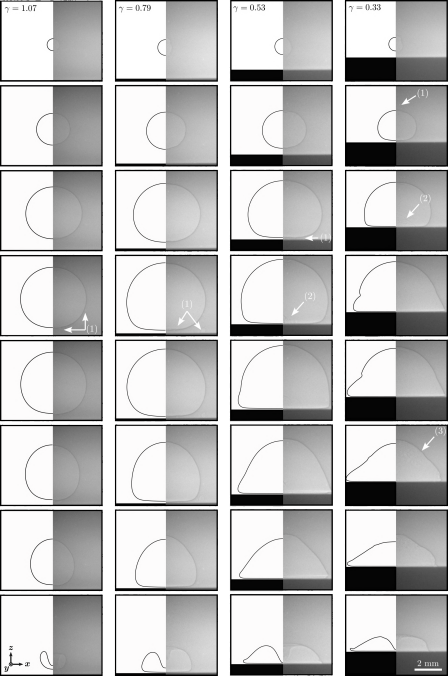
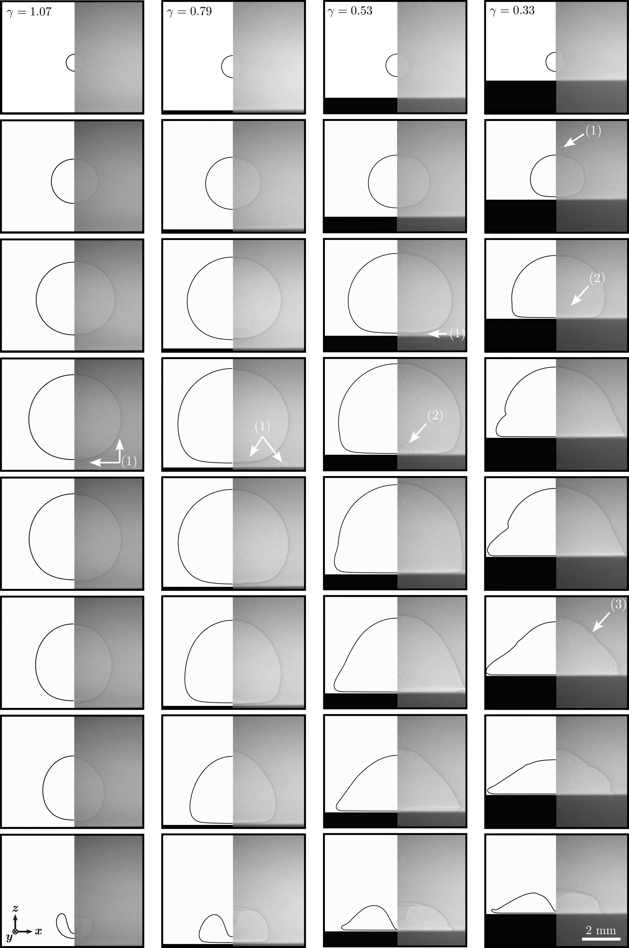
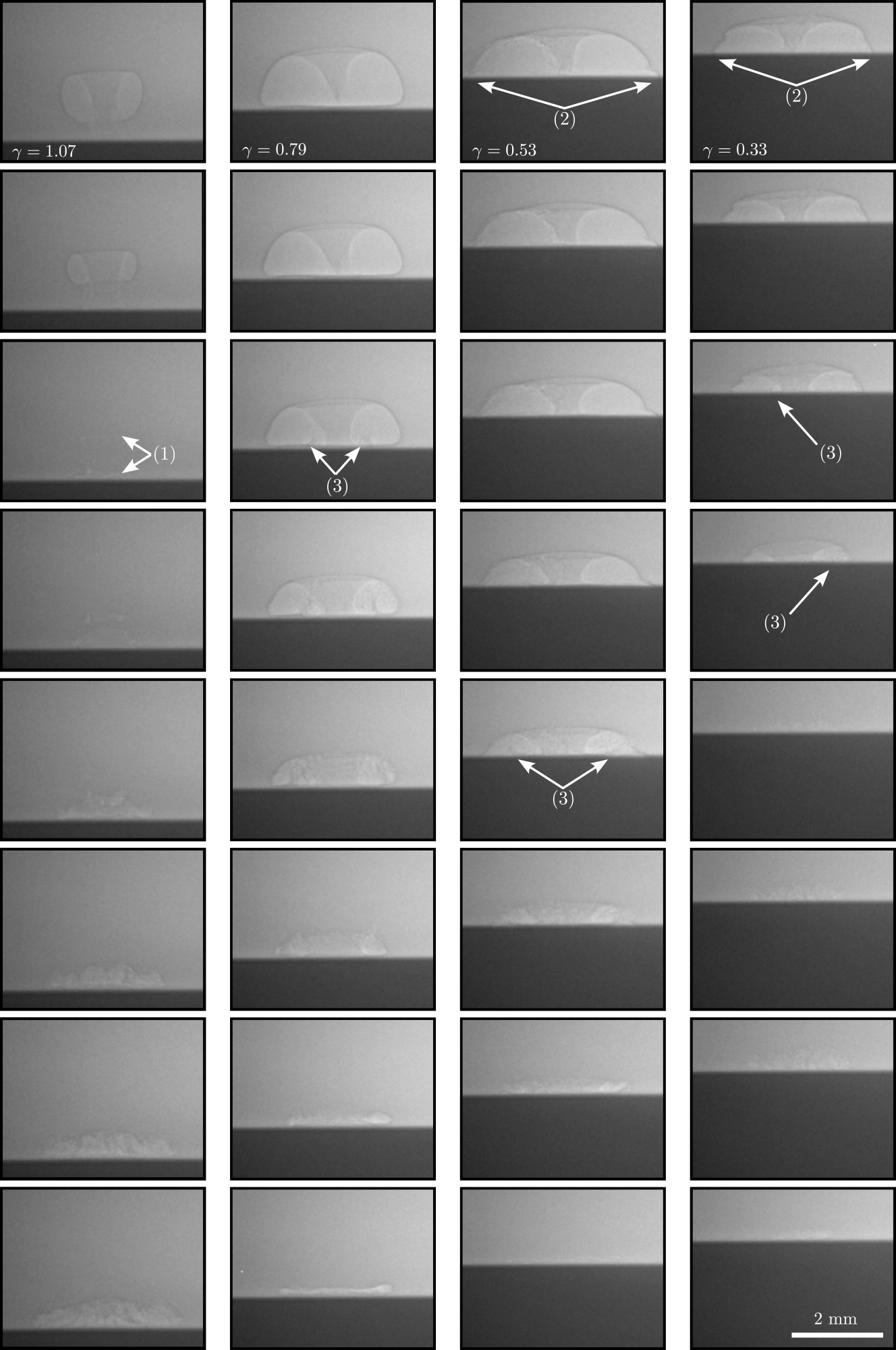
X-ray image sequences (right) and boundary integral method simulation snapshots (left) of the growth and collapse of a cavitation bubble for different stand-off parameters. The labels indicate: (1) smaller, secondary cavitation bubbles induced by the laser, (2) the ejection of a thin liquid film during the coalescence process between the main cavitation bubble and the secondary cavitation bubbles, and (3) the impact of microdroplets, originating from the breakup of the thin liquid film, against the cavity surface. -
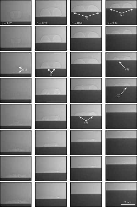
X-ray image sequences of the collapse of toroidal bubbles resulting from the impact of a liquid jet with the opposite side of a cavitation bubble for different stand-off parameters. The labels indicate (1) the bubble separation in two distinct cavities upon collapse, (2) the appearance of a protuberance, and (3) Blake’s splashing. -
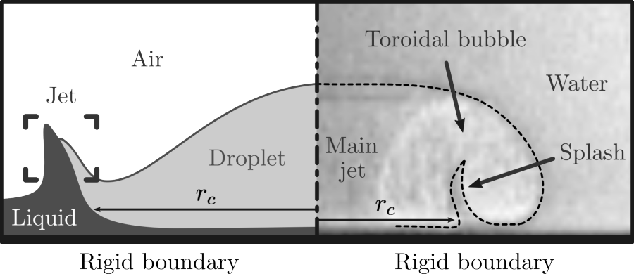
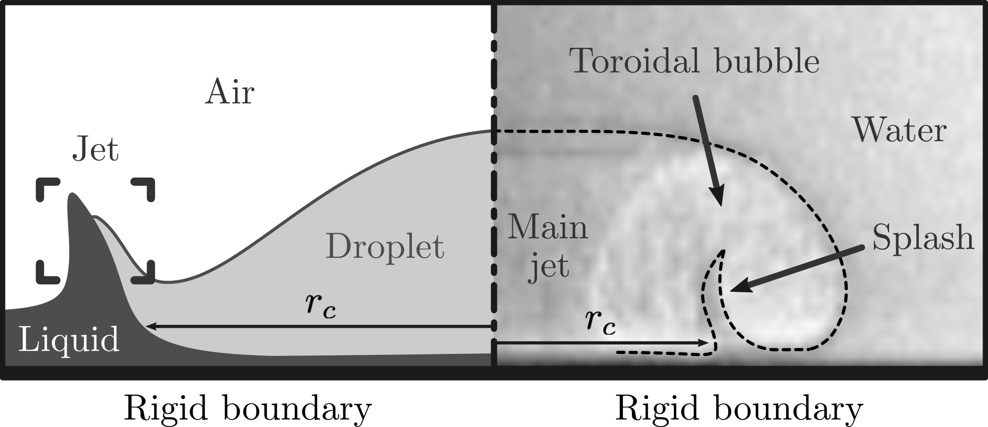
Splashing of (left) a droplet impacting on a thin liquid film (reproduced with permission from C. Josserand and S. Zaleski, Phys. Fluids 15, 1650–1657 (2003). Copyright 2003 AIP Publishing) and (right) during the collapse of a cavitation bubble near a wall. In both cases, the impact of a liquid body upon a thin liquid film results in splashing, which adopts the form of a jet evolving from an initial cusp located at rc.
Laser-induced cavitation bubble dynamics at different distances from a rigid boundary is investigated using high-speed synchrotron x-ray phase-contrast imaging (collaboration with European Synchrotron Radiation Facility external pageESRF). This is achieved through the design of a tailored experimental chamber specifically designed to reduce the x-ray absorption along the path length in water while mitigating boundary effects. The highly resolved undistorted radiographs are able to visualize a sharp bubble interface even upon complex shapes, which can serve as high-quality benchmarks for numerical simulations. Here, the measured bubble shapes are compared to simulations using the incompressible boundary integral method. The direct optical access to the high-speed liquid jet provides accurate measurements of the evolution of the jet speed, which is contrasted to the simulated results. After the jet has impacted the opposite side of the cavitation bubble, the cavity assumes a toroidal shape, the volume of which can be accurately measured from the radiographs and its temporal evolution compared to the bubble-ring model. Thanks to the clear optical access to the cavity lobes throughout the collapse, non-axisymmetric splashing within the bubble resulting from the jet impact, also known as Blake’s splashing, is observed and characterized for stand-off parameters of γ < 1. Measurements extracted from the highly resolved visualizations provided herein have been validated against scaling laws for droplet impact on a thin liquid film, which contribute to confirm and elucidate the splashing phenomenon. More details can be found in our external pagestudy.