Acoustic Droplet Vaporization
We study the phase-change process during acoustic droplet vaporization of coated microscopic droplets. Such droplets can be applied as ultrasound contrast agents or drug carriers.
Since the introduction of ultrasound as an imaging tool in 1956, its diagnostic and therapeutic applications have rapidly evolved to improve its effectiveness. However, traditional agents (microbubbles) are still too large to pass through the vascular space, thus limiting their effectiveness in targeting sites on the luminal surface of the endothelium, and often presents a low circulation time. Hence, the newest family of agents consists of droplets that are submicron or micron sized, composed by a perfluorocarbon (PFC) liquid core and a stabilizing shell made by lipid, polymer or proteins. These droplets, when exposed to ultrasound, can be vaporized into microbubbles, thus permitting their usage as ultrasound contrast agents or as drug carriers. Their small size improves their stability and permits the extravasation from leaky vasculature, features which can be greatly exploited in cancer treatment. This process is called acoustic droplet vaporization (ADV).
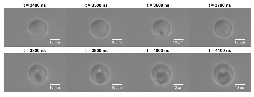
Despite their promising performance, the physics underlying the vaporization of micron-sized droplets is still to be completely discovered, while a great level of knowledge and mastery is needed in order to achieve the introduction of this technique in clinical treatment. In our laboratory we study the phase-change process, with the objective of better understanding its mechanism, discovering cutting-edge techniques to control and facilitate acoustic droplet vaporization and explore how these phenomena can improve the efficiency of ultrasound-based medical applications.